Smartphone-based medical imaging has been a flagbearer of disruptive innovation in the digital healthcare space. Owing to constant upgrades in sensors access to RAW data from sensors, and ability to do multi-threaded coding, smartphones have become the go-to imaging device for most medical practitioners, especially ophthalmologists (1). Smartphone-based devices that can screen for diseases such as cataract, infections of the anterior segment, retinal diseases, refractive error and visual field loss hold the potential to disrupt and decentralise ophthalmic screening to close-to-patient contexts, such as public health centres, primary care clinics and even in the convenience of a patient’s home, thus making eye-care more accessible and affordable.
Smartphone-based Posterior Pole Retinal imaging
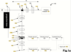
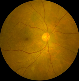
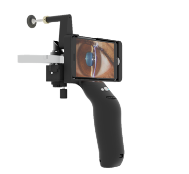
Taking the utility of conventional, desktop-based fundus cameras, one step ahead is Remidio’s FOP NM10 – the world’s first smartphone-based, patented, non-mydriatic, infrared-enabled, portable fundus imaging device that captures corneal reflex-free images of the retina with a field-of-view of 45°. It does so by using an innovative, annular illumination-based design, that separates the illumination and imaging pathways, thereby producing clear images without artefacts.

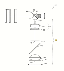
A blue tooth-based synchronization between an ISO15004 compliant LED light source and the shutter of an iPhone camera, helps produce high quality non-mydriatic images in pupils greater than 3 mm. Internal and external fixation targets help the patients focus on the required part of the device and the operator can get the best focused image on reaching the correct working distance, without pressing the trigger. These devices have been validated in multiple peer reviewed publications to produce images as good as that from Zeiss FF450 and TOPCON TRC50DX desktop retinal cameras. More than 2000 of these devices are currently being used in India, US, Singapore, Australia, Latin America, and Africa, in ophthalmology, optometry, and in public health screening. It is one of the few devices that can be used both handheld as well as on a slit lamp or on a chinrest. The device comes equipped with auto-montage – a feature that helps produce widefield images by automatically stitching multiple 45-degree field of view images.
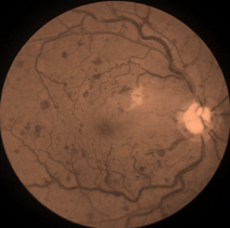
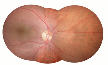
Image Courtesy: Aravind Eye Hospital, Pondicherry
The device is CE marked and US FDA510K registered. The FOP NM10 can screen for the presence of referable diabetic retinopathy (RDR) using an Offline Artificial Intelligence (AI) algorithm within a span of less than 10 seconds. The sensitivity, specificity, and efficacy of the device as a screening tool have been proven with several studies published in leading peer reviewed journals such as Nature Eye, PLOSOne, JAMA Ophthalmology and Ophthalmology Retina (2) (3) (4).
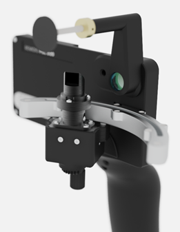
Smartphone-based Widefield Retinal imaging
A new innovation in the field of retinal imaging is Remidio’s Vistaro, a mydriatic, patented, smartphone-based, wide-field imaging device that can help screen diseases beyond the posterior pole, in a single shot. The Vistaro captures retinal images with a 65° field-of-view, with the help of external fixation targets and an automatic image-capture feature on reaching the correct working distance. The device weighs just 800 gm and can be used on a chinrest or in a handheld mode
Two images can be montaged within a span of seconds to provide an equivalent of the traditional, seven-field ETDRS image – the gold standard for early detection of retinal pathologies. Conventional wide-field and ultra-wide-field imaging devices are bulky, SLO-based that limit their use to specialized clinics and require skilled handlers. Being a smartphone-based device, Vistaro makes wide-field imaging simple and portable. Unlike traditional, non SLO based desktop fundus cameras from Zeiss and TOPCON that extend the field of view upto 50 degrees, Remidio VISTARO uses a novel optical design that extends the field of view to 60+ degrees in a single shot without compromising on the image quality.

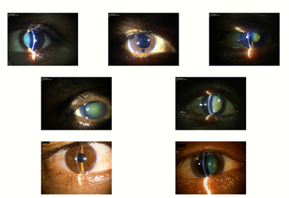
Smartphone-based Anterior Segment Imaging on Existing Slit Lamps
Remidio simplifies anterior segment imaging with the Remidio Anterior Imaging Module (AIM).
Remidio’s Anterior Imaging Module (AIM) uses high quality Anti-Reflection (AR) coated binocular optics, combined with a 50/50 beam splitter design, that matches the magnification drum optics of slit-lamps with 3-step or higher level of magnification.
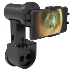
The smartphone-based design ensures instant photo documentation and the use of Apple TV, provides for instant review on large screen TV’s ensuring instant patient education.
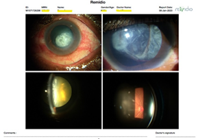
The custom app that is supplied with the AIM module ensures that the focus is determined purely by the position of the slit-lamp – i.e. it maintains a fixed focus optics, without activating the autofocus of the smartphone. Hence, the image that is viewed and captured on the smartphone is exactly what the clinician sees through the eyepiece. AIM has a universal design made to fit exactly into the smartphone sensor, requiring no additional adjustments between the eye piece and the slit-lamp. The depth of focus can be adjusted to visualise the lens, cornea, iris, or eyelids / lashes, or even the angles, as desired. AIM is used to screen for a range of anterior segment conditions. AIM also conforms to international standard, and is US FDA 510(k) exempted and CE marked. The device provides a resolution, as per Air Force Target of 56 um. The use of a Bluetooth trigger ensures that the clinician does not have to view the screen of the phone when acquiring the image.
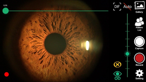
Smartphone-based Digital Portable Slit Lamp
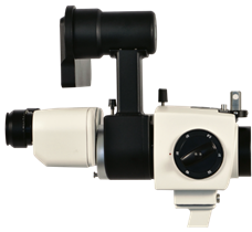
The Remidio PSL D20 is an ergonomically-designed anterior-segment screening device with an optomechanical module swivelling on a guideway, constituting the illumination system and a linear imaging module developed on a smartphone-based design. The device addresses a major challenge in anterior segment imaging using slit lamps – a small depth of focus between the planes of the iris, lens, and the cornea, by using an on-phone image processing algorithm that automatically extracts frames with peak sharpness from videos where the lens, iris and cornea may be independently in focus, to produce the best possible images. This effectively reduces, or in many cases eliminates de-focused images. The device has an adjustable headrest for convenience of (manual) focussing, that makes the object of focus visible clearly on the phone screen. This makes the device stable and allows easy capture of images just by looking at the phone screen instead of a binocular in most high-end portable slit lamps. One can effectively operate the PSL D20 using four controls: a button to toggle between background illumination lights (blue, green and white LED) and one to capture the image, a knob to adjust the slit beam-intensity; the three falling comfortably within the grasp of the thumb; a knob to adjust the variable slit-
width is located on the swivelling optomechanical unit, underneath the prism.
The controls make it convenient for the user to adjust to the ambient light conditions when imaging a patient’s eye. The optomechanical illumination unit is supported by a pair of ball spring plungers, to provide the necessary friction and prevent variation of angles when a patient is being examined. The design ergonomics make PSL D20 stand out among other portable slit lamps, while still achieving a clinical resolution of 56 um in AF Target tests.
The FOP NM10, Vistaro, AIM and PSL D20 being smartphone-based devices, are telemedicine friendly. They can be used in both synchronous and asynchronous modes of teleconsultations for ease of use and improved patient access in times like the ongoing pandemic. Their portability makes them useful even in emergency care units and on patients with limited mobility. All devices come with cloud storage on a HIPPAA compliant server for safe and secure data storage. They can be operated keeping a safe distance from patients, as may be warranted during the ongoing COVID19 pandemic.

A Zoom based Remote Consultation on Remidio AIM
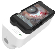
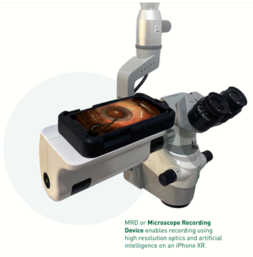
Smartphone-based 4K Wireless Video Recording:
The Microscope Recording Device (MRD) offers the convenience of wire- free, high-quality 4K recording on a smartphone-based format. It works on an intelligent acquisition software that allows for custom field of view selection, ISO control and selection of depth of focus during recording, enabling the user to create professional videos. The videos are shared to a local storage wirelessly and seamlessly using the proprietary Remidio ShareTM technology. The device can also be used to stream surgeries live on third party applications like YouTube and Facebook, in full HD or 4K.
The use of screen mirroring with Apple TV allows for the images and videos captured on Remidio’s smartphone-based devices, to also be seen with minimal lag wirelessly on Smart TVs for the education of the patient along with training of residents.
The device has been designed ergonomically to meet the eye level of the doctors during surgery. The device comes with a Windows PC based patient video management software, with the ability to edit the videos and insert voice over and annotations.
Conclusions:
A smartphone-based user interface is easy to use and the devices can be operated after basic training by non-specialist operators with minimal supervision. Hence, such devices offer benefits of being scalable solutions to screen for diseases responsible for preventable blindness in remote areas with limited access to specialists. Telemedicine helps provide instant consultations from specialists and helps triage patients in cases of emergencies. Innovations like these unleash the tremendous potential of telemedicine-based screening system and have the ability to effectively reduce the global burden of preventable blindness.
References:
- Chhablani J, Kaja S, Shah VA. Smartphones in ophthalmology. Indian J Ophthalmol. 2012;60(2):127–31.
- Sosale B, Aravind SR, Murthy H, Narayana S, Sharma U, Gowda SGV, et al. Simple, Mobile-based Artificial Intelligence Algo r ithm in the detection of Diabetic Retinopathy (SMART) study. BMJ Open Diabetes Res Care. 2020 Jan;8(1):e000892.
- Natarajan S, Jain A, Krishnan R, Rogye A, Sivaprasad S. Diagnostic Accuracy of Community-Based Diabetic Retinopathy Screening With an Offline Artificial Intelligence System on a Smartphone. JAMA Ophthalmol. 2019 Oct 1;137(10):1182–8.
- Sengupta S, Sindal MD, Baskaran P, Pan U, Venkatesh R. Sensitivity and Specificity of Smartphone-Based Retinal Imaging for Diabetic Retinopathy. Ophthalmol Retina. 2019 Feb;3(2):146–53.