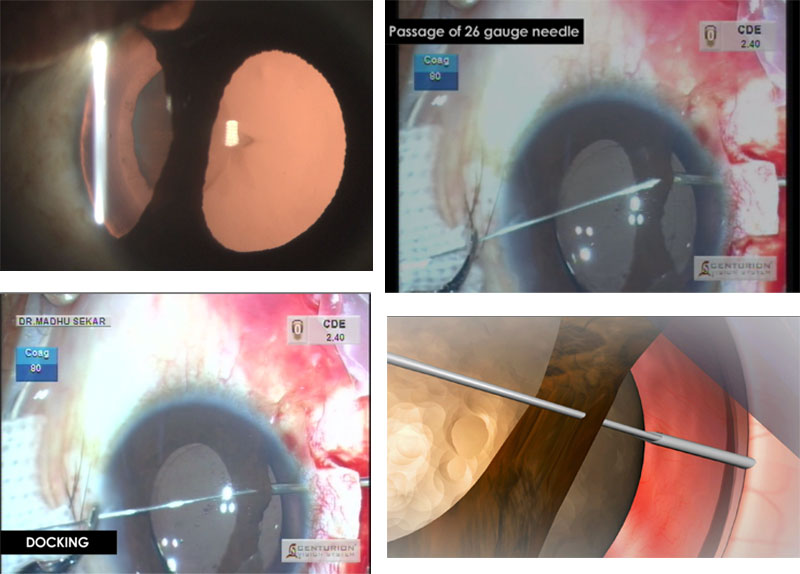
Iridodialysis occurs following blunt trauma, penetrating trauma and during cataract surgery. Here are two videos showing, traumatic cataract extraction with iridodialysis repair (following blunt trauma) along with the animation of the repair. Phaco-emulsification with iridodialysis repair was done in both cases. Procedure: Conjunctival peritomy was done near the iridodialysis site. Limbal based partial thickness scleral flap created alongside the iridodialysis. Paracentesis was made opposite to the iridodialysis site. A 26 gauge needle was inserted under the sclera flap, 1.5mm behind the limbus, into the anterior chamber. A double-armed, straight needle, 10-0 polypropylene suture, was passed through the iris root from paracentesis, docked to the 26 gauge needle and pulled out. The process was repeated with the other arm. The shape of the pupil was assessed and suture tightened with 5-6 knots under the sclera flap. Conjunctiva was closed with 8-0 vicryl suture. Quality of vision is as important as quantity of vision
By,
Dr Madhu Sekar