Field of ophthalmology is ever-evolving with newer advances technologically as well as diagnostically.
Covid-19 has brought many landmark changes in almost every field of medical science as well as various day-to-day activities.
Ophthalmology is also a part of these newer advances as an adaptation to Covid-19 response.
Corneal transplants no more hold challenge in this new age, but Covid era brought a significant change, as there has been a dearth of tissues, considering the scare of spread of infection from the donor to the recepient.
In the following segment, we shall be discussing few advancements in the field of cornea and refractive services.
1. Microbiology
Novel use of trypan blue to stain the fungal hyphae in corneal scrapings in eye clinics.
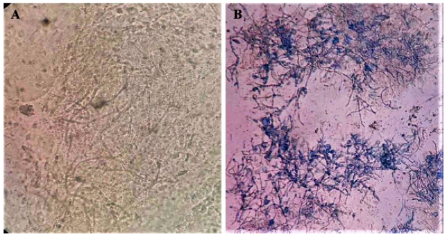
Hyaline filaments (A) and with trypan blue dye staining (B) showing bluish-colored filaments
USES OF TRYPAN BLUE STAINING OF FUNGAL HYPHAE –
- Highlights the fungal hyphae for detection by untrained eyes as well
- Useful as a teaching tool
- Enhances the smartphone photography
- Aids in teleophthalmology, as highlights the fungal hyphae better from the surroundings which can be easily shared electronically as well.
2. Collagen cross-linking techniques
Collagen cross-linking (CXL) for thin corneas can be enhanced by:
- DSAEK-Lenticule assisted CXL, b. contact-lens assisted CXL , c. Corneal cross-linking (CXL) combined with refractive surgery (CXL plus)
- Slit-lamp based CXL – C-eye device
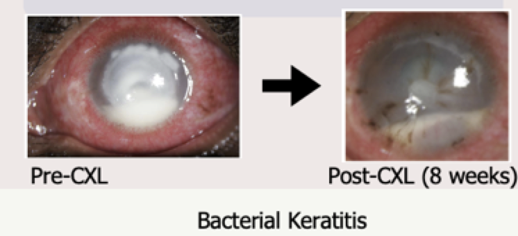
- Cross linking in infectious keratitis patients
- CXL in management of advanced non-resolving microbial keratitis – Cxl can be used in management of superficial microbial keratitis and as an adjunctive treatment in the management of advanced non-resolving keratitis
3. INFECTIOUS KERATITIS
Rose Bengal Photodynamic antimicrobial therapy (RB-PDAT) for patients with progressive infectious keratitis.4
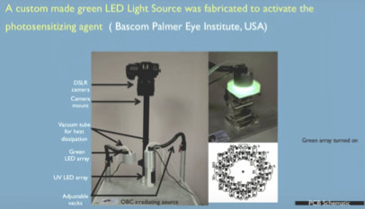
RB-PDAT can be considered as an adjunct for management of severe, progressive infectious keratitis before considering for therapeutic keratoplasty.
4. CORNEAL TRANSPLANTATION
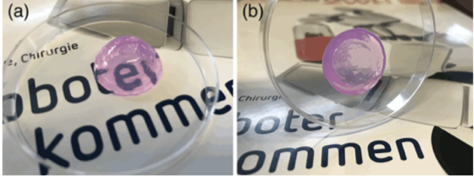
A) Corneal bio-prints: Transparent bioprinted dome-shaped artificial corneas consisting of 0.5% agarose with 0.2% collagen Type I can be used clinically for patients with corneal stromal diseases.
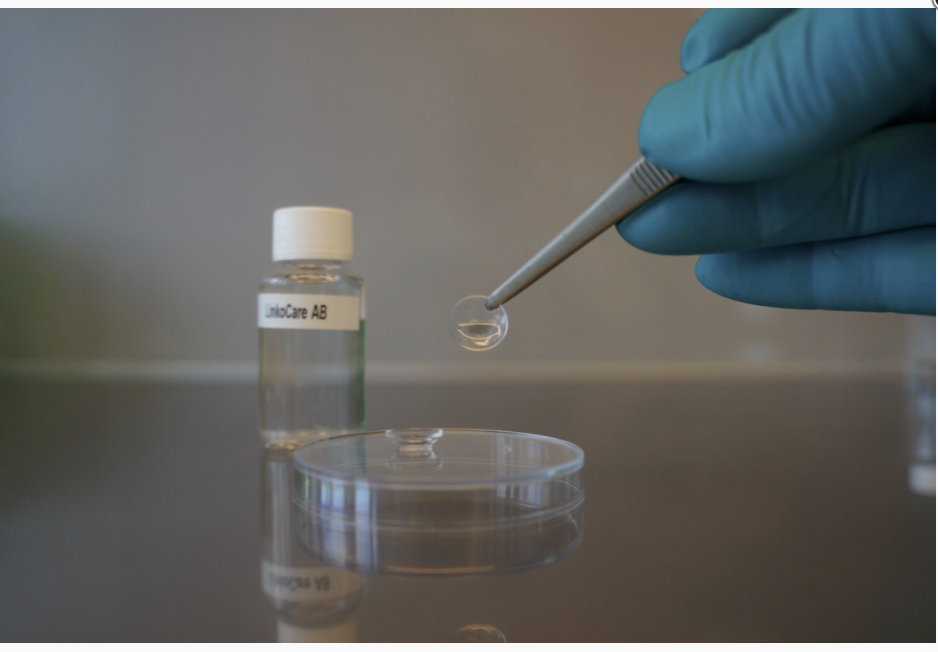
B) Sutureless Femtolaser-assisted anterior lamellar keratoplasty using a bioengineered cornea (linkcor) as a viable alternative to human donor transplantation for superficial corneal opacities.
5. Treatment of endothelial diseases
A) Rho-kinase inhibitors (ROCK inhibitors) – ROCK inhibitor eyedrops can promote proliferation of residual corneal endothelial cells and increase the number of corneal endothelial cells for coverage , thereby reducing the risk of corneal decompensation.7
They can be used to treat corneal endothelial diseases post-cataract surgery or Fuch’s endothelial dystrophy.

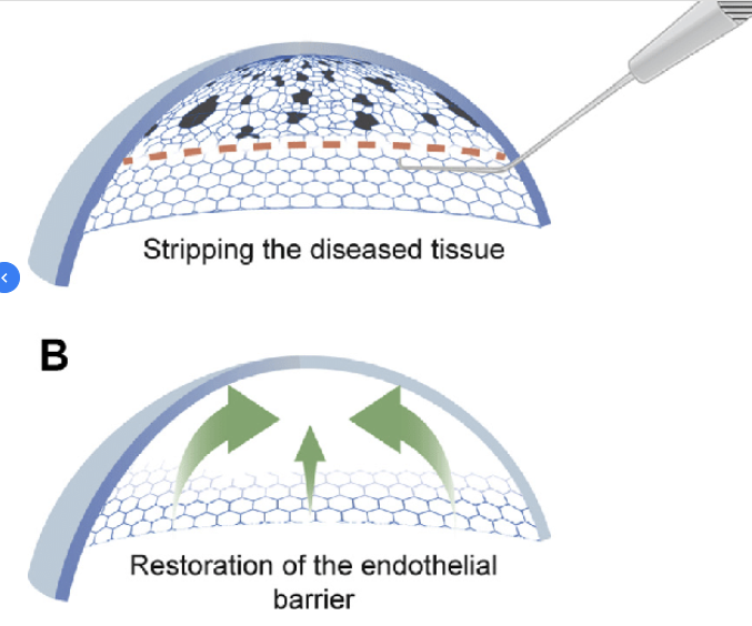
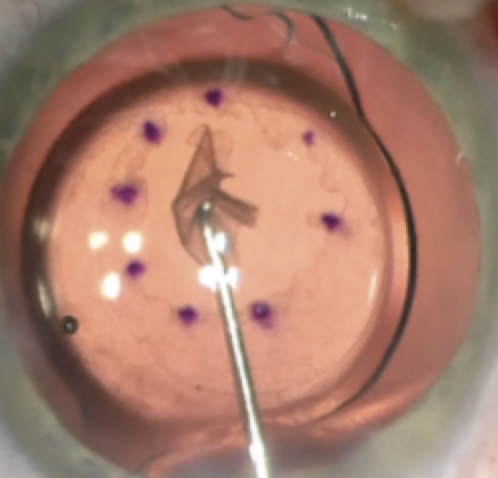
B) Descemet’s stripping only for a chronic descemet’s detachment after cataract surgery –8 Descemet’s stripping only for a chronic descemet’s detachment after cataract surgery – Descemet’s stripping only is gaining acceptance for corneal endothelial decompensation in Fuch’s dystrophy patients as well. Addition of Rho-kinase inhibitors improves the predictability, but further trials are required to understang longer term outcomes.
Refractive Surgery
- PresbyLASIK – Lasik with aspheric ablation profiles and a micro-monovision protocol is an effective option for presbyopia correction in pseudophakic patients. Other treatment modalities are PresbyMAX, Supracor, and laser blended vision.
- SMILE Xtra – Corneal cross-linking combined with SMILE procedure (SMILE Xtra) is a promising tool to prevent ectasia in high-risk patients. It has proven to be safe and simple procedure offered to patients undergoing SMILE with risk for ectasia.
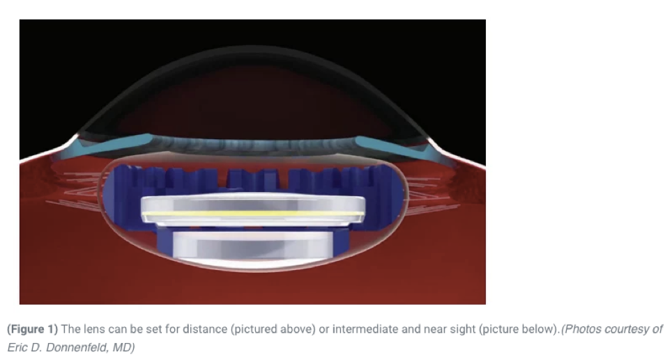
- Curvature changing IOLs – This novel IOL comprises a base lens supported by circumferential silicone haptics that fill the capsule and a fluid lens that fits into the base lens and facilitates continuous range of vision from distance to near focus.
AUTHOR
Dr. Gunjan Budhiraja,
Cornea and Refractive surgeon
Dr Shroff’s Eye Hospital – Delhi, INDIA
REFERENCES:
1.Prasher P, Singh B, Singh K. Novel Use of Trypan Blue Dye to Stain Fungus in Corneal Scrapings in Eye Clinics. Cornea. 2020 Nov;39(11):e26. doi: 10.1097/ICO.0000000000002446. PMID: 32769676.2. https://www.emagine-eye.com/slit-lamp-cxl/
3. Shetty R, Nagaraja H, Jayadev C, Shivanna Y, Kugar T. Collagen crosslinking in the management of advanced non-resolving microbial keratitis. Br J Ophthalmol. 2014 Aug;98(8):1033-5. doi: 10.1136/bjophthalmol-2014-304944. Epub 2014 Apr 7. PMID: 24711659.
4. Naranjo A, Arboleda A, Martinez JD, Durkee H, Aguilar MC, Relhan N, Nikpoor N, Galor A, Dubovy SR, Leblanc R, Flynn HW Jr, Miller D, Parel JM, Amescua G. Rose Bengal Photodynamic Antimicrobial Therapy for Patients With Progressive Infectious Keratitis: A Pilot Clinical Study. Am J Ophthalmol. 2019 Dec;208:387-396. doi: 10.1016/j.ajo.2019.08.027. Epub 2019 Sep 5. PMID: 31493402; PMCID: PMC7184264.
5.Duarte Campos DF, Rohde M, Ross M, et al. Corneal bioprinting utilizing collagen-based bioinks and primary human keratocytes. J Biomed Mater Res. 2019; 107A:1945–1953. https://doi.org/10.1002/jbm.a.36702
6. Khodaparast M, Shahraki K, Jabbarvand M, Shahraki K, Rafat M, Moravvej Z. Sutureless Femtosecond Laser-Assisted Anterior Lamellar Keratoplasty Using a Bioengineered Cornea as a Viable Alternative to Human Donor Transplantation for Superficial Corneal Opacities. Cornea. 2020 Sep;39(9):1184-1189. doi: 10.1097/ICO.0000000000002394. PMID: 32558727.
7. Okumura N, Kinoshita S, Koizumi N. Application of Rho Kinase Inhibitors for the Treatment of Corneal Endothelial Diseases. J Ophthalmol. 2017;2017:2646904. doi: 10.1155/2017/2646904. Epub 2017 Jul 2. PMID: 28751979; PMCID: PMC5511675.
8. Hirabayashi KE, Mark D, Lau J, Lin CC. Descemet Stripping Only for a Chronic Descemet Detachment After Cataract Surgery. Cornea. 2020 Mar;39(3):379-381. doi: 10.1097/ICO.0000000000002195. PMID: 31725698.
9. Garcerant D, Hirnschall N, Toalster N, Zhu M, Wen L, Moloney G. Descemet’s stripping without endothelial keratoplasty. Curr Opin Ophthalmol. 2019 Jul;30(4):275-285. doi: 10.1097/ICU.0000000000000579. PMID: 31033737.
10. Elmohamady MN, Abdelghaffar W, Bayoumy ASM, Gad EA. Correction of pseudophakic presbyopia using Lasik with aspheric ablation profiles and a micro-monovision protocol. Int Ophthalmol. 2021 Jan;41(1):79-86. doi: 10.1007/s10792-020-01554-7. Epub 2020 Sep 9. PMID: 32902784.
11. Shetty R, Brar S, Sharma M, Dadachanji Z, Lalgudi VG. PresbyLASIK: A review of PresbyMAX, Supracor, and laser blended vision: Principles, planning, and outcomes. Indian J Ophthalmol. 2020 Dec;68(12):2723-2731. doi: 10.4103/ijo.IJO_32_20. PMID: 33229648; PMCID: PMC7857007.
12. Osman IM, Helaly HA, Abou Shousha M, AbouSamra A, Ahmed I. Corneal Safety and Stability in Cases of Small Incision Lenticule Extraction with Collagen Cross-Linking (SMILE Xtra). J Ophthalmol. 2019 Apr 14;2019:6808062. doi: 10.1155/2019/6808062. PMID: 31098325; PMCID: PMC6487093.
13. https://www.ophthalmologytimes.com/view/curvature-changing-iol-new-frontier-in-cataract-and-presbyopia-surgeries