It is not often that an eye surgeon is able to awe a room full of medical students and residents in a busy Ophthalmology clinic. So I’ll take this opportunity to write the story about this this extra-ordinary accomplishment.
An 18yr old male patient was referred to me from elsewhere. The patient came with complaints of a swelling on his left eyebrow since last 3 months. He had gone to several doctors of different specialties before me and had several provisional diagnosis ranging from Sebaceous Cyst to Schwannoma to Dermoid cyst!
At first look it did seem to be that one of those diagnoses was right. On examination though, the texture felt like the classical “bag of worms” sign of a particular condition. I asked for history including leading questions of specific signs which would confirm what I suspected. All negative!

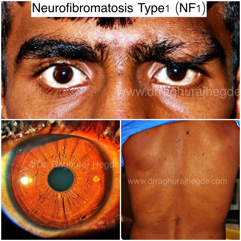
The clincher was by the slit lamp! Lisch Nodules-yellow to brown coloured melanocytic harmartomas- dome shaped projecting out of iris which are harmless and don’t affect vision. However they are present in up to 94% of Neurofibromatosis type 1.

I repeated my questions about other signs of NF1. Café au lait spots-flat brown medium-large skin spots in the body. 6+ spots is strongly indicative of NF1. Neurofibromas- Small pea-sized bumps on the skin mostly at the back. All denied again!
However, I was confident enough to ask him to lift his shirt so that I could examine his back. I think I heard a gasp of awe behind me from the medical students/interns/residents who were watching.

The café au lait spots and few neurofibomas spread across his back.
NF1 confirmed. Patient advised for imaging to exclude other features of NF1. Also advised for excision of left brow plexiform neurofibroma with brow-pexy to correct the cosmetic blemish and restore symmetry of the face
Will post this patient’s after surgery picture soon!
©All patient photos are being used with the express consent of the patient. These cannot be shared or reproduced elsewhere.
Additional Information
Neurofibromatosis is caused by genetic mutations that either are passed on by a parent or sometimes occur spontaneously during pregnancy. The specific genes involved depend on the type of neurofibromatosis:
- NF1. The NF1 gene is located on chromosome 17. This gene produces a protein called neurofibromin that helps regulate cell growth. The mutated gene causes a loss of neurofibromin, which allows cells to grow uncontrolled.
- NF2. The NF2 gene is located on chromosome 22, and produces a protein called merlin (also called schwannomin), which suppresses tumors. The mutated gene causes a loss of merlin, leading to uncontrolled cell growth.
- Schwannomatosis. So far, two genes are known to cause schwannomatosis. Mutations of the genes SMARCB1 and LZTR1, which suppress tumors, are associated with this type of neurofibromatosis.
Published by Dr. Raghuraj Hegde