Smartphone photography has become essential in Ophthalmic practice and a standard way of recording ocular findings for documentation, expert opinion, academics, tele-ophthalmology and patient education. Anterior segment photography has been made possible using adapters which help fix the smartphone to the slit lamp or adapters with lens which can be fixed to the phone camera that are commercially available.
The need for a simpler instrument to view and record anterior segment finding is prudent for any ophthalmologist. Anterior segment photography with Intraocular lens(ASPI) is one such instrument which can help any ophthalmologist take anterior segment photos on the go without the help of slit lamp.
Preparing the ASPI
- 2 strips of 4 cm × 2 cm hard chart paper/plastic sheet were cut
- A circular opening of 5 mm diameter was made on both the strips at one end using a No. 11 surgical blade
- The intraocular lens was placed on the opening of one strip by applying liquid adhesive over the haptics of the IOL (alternatively, double-sided tape can also be used to fix the IOL and the second strip in place)
- The second strip was then stuck on top of the IOL and the first strip so as to cover it
- This arrangement was then aligned on the smartphone camera with a Micropore/Cellotape
- The smartphone was then moved towards the patient to focus on the lesion and a still photograph or a video was recorded with the inbuilt flash or an external light source (torch)
- The images obtained were of high quality with high magnification and moreover, it is a device that can be made using materials available in the clinic/hospital
Smartphones are beginning to play a major role as a medical diagnostic tool. They are simple, stable, affordable as well as being portable, with high storage capacity and wireless connectivity makes the ASPI an important tool for diagnosis and documentation in various setups such as clinics, hospitals, health centers, and camps.
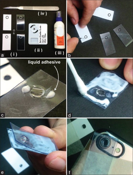
figure 1 : (a) Materials required to prepare the ASPI: (i). Hard chart paper/Plastic strip: 4 cm × 2 cm (ii). IOL (iii). Liquid adhesive (iv). No. 11 Surgical blade (b) Making the 5 mm circular hole on the strip using the no. 11 surgical blade. (c) Fixing the IOL on the circular hole by applying liquid adhesive on the haptics. (d) and (e) Fixing the second strip on the first strip and IOL using adhesive. (f) Placing the ASPI on the Smartphone camera
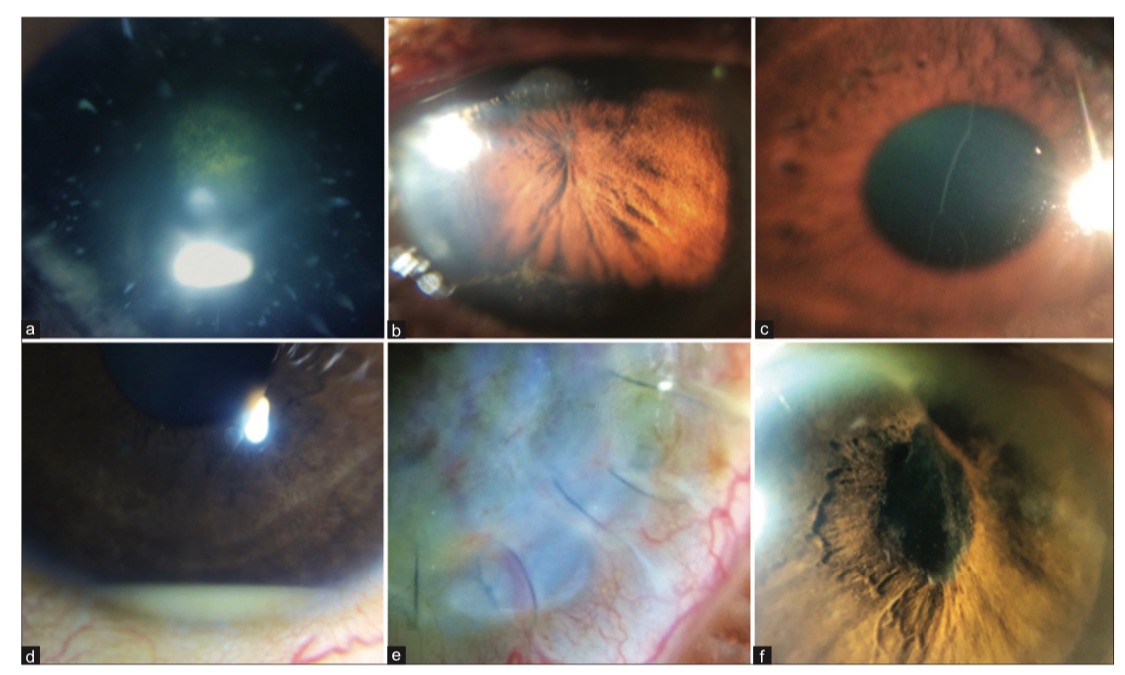
figure 2 : (a) Blue dot cataract with Posterior subcapsular cataract. (b) Occlusio pupillae. (c) Prominent corneal nerve. (d) Hypopyon. (e) Keratoplasty sutures. (f) Pupillary fibrin membrane
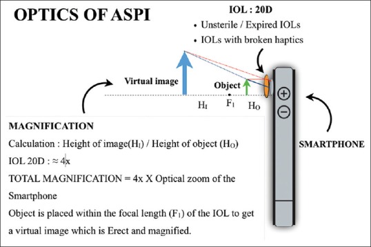
figure 3 : Optics of ASPI (Anterior Segment Photography with Intraocular lens)
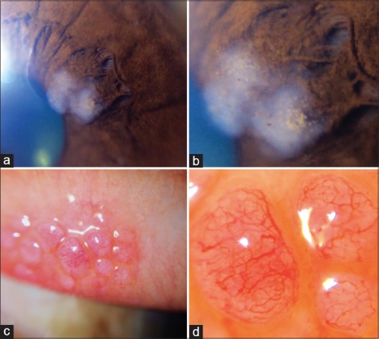
figure 4 : (a) Iris mass: 4× zoom (b) Iris mass: 30× zoom (c) Giant papillae: 4× zoom (d) Giant papillae: 30× zoom
AUTHOR
Dr. Prithvi Chandrakanth,
Aravind Eye Hospital,
Coimbatore