Predicting Prognosis Based on Regional Prevalence, Ulcer Morphology and Treatment Strategy in Vision-Threatening Pythium insidiosum Keratitis.
Clin Ophthalmol. 2023 May 5;17:1307-1314. doi: 10.2147/OPTH.S412274. PMID: 37181081; PMCID: PMC10167989.
Author – Gurnani B, Kaur K. MBBS (AFMC PUNE), DNB (AECS), FCRS (AECS), FICO(UK), FAICO (Ref Sx), MRCS Ed (Ophthal), MNAMS (NEW DELHI) Former Consultant- Department of Cataract, Cornea and Refractive, AECS, Pondicherry, India, Dr. Om Parkash Eye Institute, Punjab, Amritsar, India
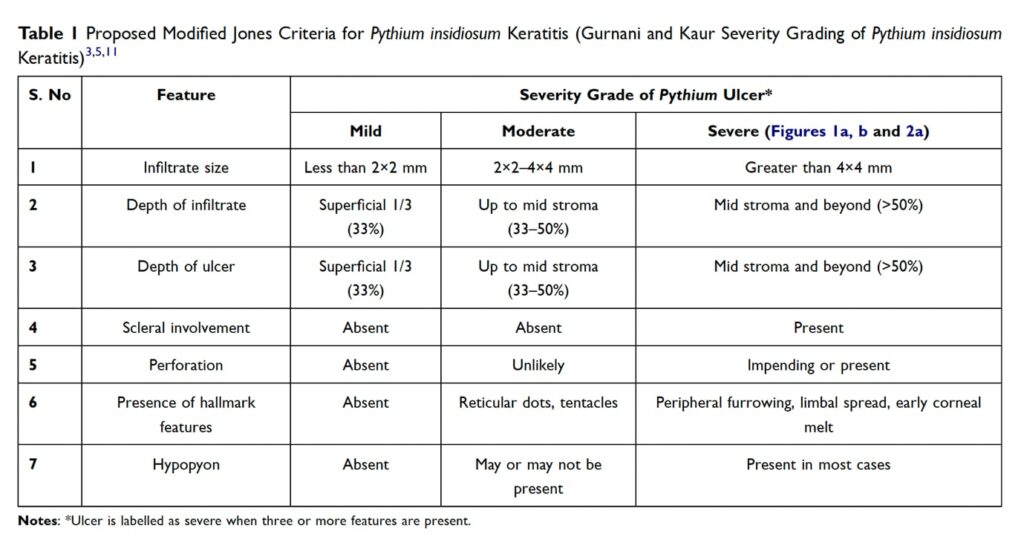
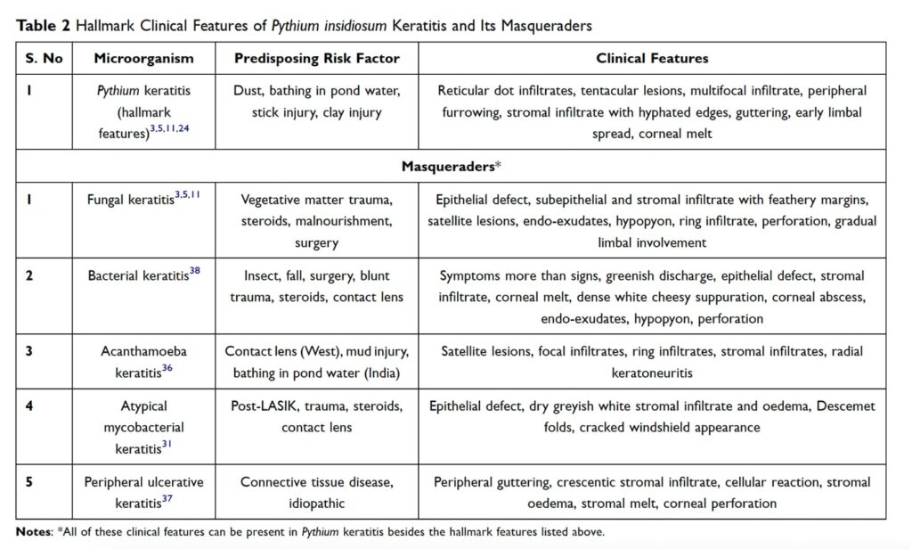
Pythium insidiosum is an oomycete residing in aquatic bodies, with a higher prevalence in tropical and subtropical regions. It causes vision-threatening keratitis and closely mimics fungal keratitis, making it a “parafungus.” The first case of systemic pythiosis was reported in Thailand in 1884, and the first case of keratitis was reported in 1988. The prognosis of Pythium keratitis is dependent on the regional prevalence of the species, corneal infiltrate size, location, and density at presentation, and initial treatment strategy. The classical hallmark features of Pythium include reticular dots, tentacles, hyphated edges, peripheral furrowing, and early limbal spread. The laboratory diagnosis is made by microbiological sampling and culture on nutritional agar and zoospore identification by the leaf incarnation method. The time-tested recommended treatment modalities are antifungals (5% natamycin, 1% itraconazole, 1% voriconazole), antibacterials (0.2% linezolid, 1% azithromycin, 4% minocycline, and 1% tigecycline), and therapeutic keratoplasty. However, treatment protocols vary from center to center, and specific antimicrobial therapy remains undetermined. To safeguard patients from this vision-threatening entity, the prognosis of Pythium keratitis is dependent on the regional prevalence of the species, the corneal infiltrate size, location and density at presentation, and initial treatment strategy. The presence of hallmark clinical features, such as early limbal spread, peripheral guttering, furrowing, and tentacular projections, provides an indicator of the severity and virulence of microorganisms and usually has poorer visual outcomes.
The proposed treatment strategies for Pythium include antifungals, antibacterials, therapeutic keratoplasty, cryotherapy with ethanol to the host bed, cyanoacrylate glue with bandage contact lens, enucleation, and evisceration. Antibacterials should be the first line drugs in culture-proven Pythium cases, and antifungals still have a role to play if the diagnosis is delayed. The literature review and day-to-day clinical experience suggest that the prognosis of this vision-threatening keratitis is governed by regional prevalence, infiltrate size, density, and location at presentation, and initial medical management. Pythium keratitis is a common eye condition characterized by ulcer morphology, visual axis involvement, and recurrent graft infection. The prognosis of Pythium keratitis cases in Thailand, China, Israel, and Australia is worse than that of those from India, likely due to differences in regional prevalence and virulence of the species. The Jones criteria have been proven to be poor prognostic factors for Pythium keratitis, with ulcer size, visual axis involvement, mid-to-posterior stromal infiltrate, endo-exudates, corneal perforation, early limbal spread, scleritis, and endophthalmitis being poor predictive factors. The treatment strategy for Pythium keratitis has been analyzed, with Hou et al. showing that ulcers worsened after initial treatment with antifungals, and all patients underwent therapeutic keratoplasty. Gurnani et al. emphasized that antifungals should be initiated before culture results are known, and antibacterials should be initiated once culture positivity of Pythium has been confirmed. Antibacterials have been shown to have a higher efficacy than antifungals, and Gurnani et al. successfully managed a 9-year-old paediatric patient with cyanoacrylate glue, topical linezolid, and azithromycin. This comprehensive literature review reveals that prompt diagnosis and targeted treatment can salvage these eyes. Hallmark clinical features of Pythium keratitis, along with its masqueraders, are essential for ophthalmologists and cornea specialists to understand and manage the condition. A diagnostic and management protocol can be developed to safeguard the anatomical and functional outcomes in these cases.
Pythium insidiosum keratitis diagnosis and treatment remain challenging for ophthalmologists due to the mimicry of fungal filaments. Antifungals and antibacterials have shown better outcomes in culture-proven cases. A new diagnostic and treatment algorithm is proposed for Pythium insidiosum keratitis, which has not been previously described. The algorithm aims to identify hallmark features, consider masqueraders, and use early therapeutic keratoplasty for rapidly proliferating cases. The proposed algorithm will help manage cases with good anatomical and functional outcomes.